Overview
The scanning electrochemical microscope (SECM) was introduced in 19891 as an instrument that could examine chemistry at high resolution near interfaces. By detecting reactions that occur at a small electrode (the tip) as it is scanned in close proximity to a surface, the SECM can be employed to obtain chemical reactivity images of surfaces and quantitative measurements of reaction rates. Numerous studies with the SECM have now been reported from a number of laboratories all over the world, and the instrument has been used for a wide range of applications, including studies of corrosion, biological systems (e.g., enzymes, skin, leaves), membranes, and liquid/liquid interfaces.2,3 Trapping and electrochemical detection of single molecules with the SECM has also been reported.
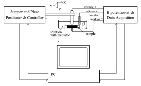
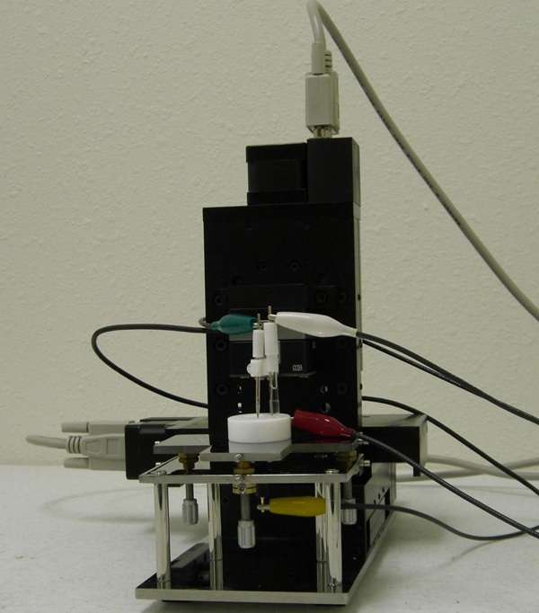
The CHI920D Scanning Electrochemical Microscope consists of a digital function generator, a bipotentiostat, high resolution data acquisition circuitry, a three dimensional nanopositioner, and a sample and cell holder. Diagrams for the SECM and cell/sample holder are shown below. The three dimensional nanopositioner has a spatial resolution down to nanometers but it allows a maximum traveling distance of 50 millimeters. The potential control range of the bipotentiostat is ± 10 V and the current range is ± 250 mA. The instrument is capable of measuring current down to sub-picoamperes.
In addition to SECM imaging, other modes of operation are available for scanning probe applications: Probe Scan Curve, Probe Approach Curve, Surface Interrogation SECM, and Surface Patterned Conditioning. Probe Scan Curve mode allows the probe to move in the X, Y, or Z direction while the probe and substrate potentials are controlled and currents are measured. The probe can be stopped when the current reaches a specified level. This is particularly useful for searching for an object on the surface and determining approach curves. Probe Approach Curve mode allows the probe to approach the surface of the substrate. It is also very useful for distinguishing the surface process using PID control. The step size is automatically adjusted to allow fast surface approach, without letting the probe touch the surface. Surface Patterned Conditioning allows the user to edit a pattern for surface conditioning by controlling the tip at two different potentials and durations. Constant height, constant current, potentiometric, and impedance modes are available for SECM imaging and probe scan curve.
The 920D is designed for scanning electrochemical microscopy, but many conventional electrochemical techniques have also been integrated for convenience, such as CV, LSV, CA, CC, DPV, NPV, SWV, ACV, SHACV, FTACV, i-t, DPA, DDPA, TPA, SSF, STEP, IMP, IMPE, IMPT, and CP. When the instrument is used as a bipotentiostat, the second channel can be controlled at an independent constant potential, to scan or step at the same potential as the first channel, or to scan with a constant potential difference with the first channel. The second channel works with CV, LSV, CA, DPV, NPV, DNPV, SWV, and i-t.
The 920D SECM is an upgrade to the 900/910B/920C SECM. The 920D uses a stepper motor positioner in conjunction with a closed-loop 3-dimensional piezo positioner. The stepper motor positioner has a resolution of 8 nanometers with 50 mm travel distance. Closed-loop piezo control allows improved linearity and reduced hysteresis of the piezo devices. Improvements include very stable and accurate potential and current control, and dual-channel data acquisition at high speed (1 MHz with 16-bit resolution).
1. A. J. Bard, F.-R. F. Fan, J. Kwak, and O. Lev, Anal. Chem. 61, 132 (1989); U.S. Patent No. 5,202,004 (April 13, 1993).
2. A. J. Bard, F.-R. Fan, M. V. Mirkin, in Electroanalytical Chemistry, A. J . Bard, Ed., Marcel Dekker, New York, 1994, Vol. 18, pp 243-373.
3. A. J. Bard and M. V. Mirkin, Eds. Scanning Electrochemical Microscopy, Marcel Dekker, New York, 2001.